
Tridactyls vs Humans
A comprehensive scientific study comparing the unique biological structures of Tridactyl specimens with modern Human anatomy using advanced CT imaging.
Pictorial essay © K H Fung / Tridactyls.org
Human-Like Tridactyls vs. Humans Comparative Report
Methodology
- Specimens included: Maria (aged adult, uncertain sex with Y chromosome); Montserrat (young pregnant female); Sebastian (adolescent).
- Preservation: Desiccated corpses with internal organs, tendons, coprolites with seeds, brain remnants, and metallic implants fused with tissue.
- Advanced CT Techniques: Including MPR, MIP, VR3D, SSD 3D, semi-transparency, and false colors. Overcame artifacts from metal and diatomaceous earth.
Featured Specimens in this Comparative Study
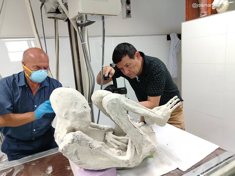
Montserrat
Dr. Zuniga and Jois Mantilla documenting the young pregnant specimen Montserrat during a detailed physical and imaging review.
Learn more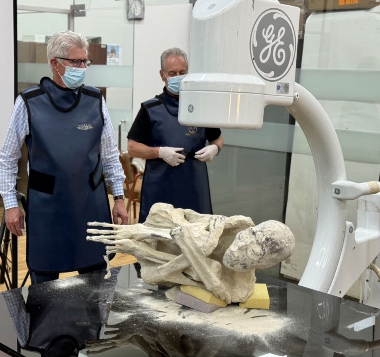
Maria
Forensic Specialist Dr. John McDowell leads an international team in the examination of the Maria specimen.
Learn more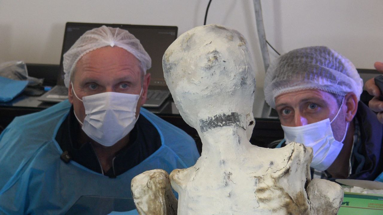
Sebastian
Investigative reporter Ross Coulthart and J.P. Hague observing the adolescent specimen Sebastian for NewsNation.
Learn moreAnatomical Similarities
| Feature | Tridactyls | Human Comparison |
|---|---|---|
| Long Bones | Paired (radius/ulna, tibia/fibula) | Standard human arrangement |
| Joints | Elbows/knees with patellae, shoulders, hips ball-socket | Functionally and structurally similar |
| Sacro-iliac | Clear sacro-iliac joints | Present in humans |
| Skull Base | Vascular markings | Similar biological blueprint |
| Dental | Grossly similar tooth structure | Similar morphology |
| Internal Organs | Remnants of heart, vessels, diaphragm, bowels, bi-lobed brain | Similar morphology organ systems |
Anatomical Differences
| Feature | Tridactyls | Human Comparison |
|---|---|---|
| Skull | Early suture fusion, posterior elongation | Sutures are present, rounder shape |
| Orbits/Face | Large, sloping, widely separated orbits | Forward-facing, closer orbits |
| Nose/Ears | Variable or absent pinnae | Prominent nose and external ears |
| Hands | 3 digits, 5 segments each, no palm | 5 digits, 3 segments, metacarpal palm |
| Feet | 3 digits, 4 segments each, claw-like, no metatarsals | 5 digits, tarsal/metatarsal structure |
| Spine | Coccyx not identified in ‘Montserrat’; identifiable in ‘Sebastian’; absent in ‘Maria’ due to sacral trauma, calcified disks in young specimens | Coccyx present, disks calcify with age/disease |
| Thorax/Abdomen | Pectus excavatum, no umbilicus, off-center uterus | Normal Thorax, pectus excavatum is a normal variant, umbilicus present |
| Implants | Fused metallic implants over hair tissue and intra-osseous (Maria) | Not naturally occurring |
Conclusion
CT imaging proves sophisticated biological structures, dispelling hoax claims. There is firm evidence of an unknown bipedal hominid species.
- Sophisticated biological structures indicate authenticity.
- No evidence of surgical manipulation or fabrication found.
- Unknown bipedal hominid species with distinct anatomical features.
- Authentic artifacts suggest historical presence.
Research Document
Core Findings
- Evidence of real biological entities — unknown bipedal hominid species, not a hoax
- CT imaging proves sophisticated biological structures, dispelling hoax claims. There is firm evidence of an unknown bipedal hominid species.
Note: The people pictured in this study are involved in the scientific analysis and documentation of the specimens. They are not the authors of the current pictorial essay.
Dr. K.H. Fung
Retired Radiologist (40+ years experience in diagnostic radiology and 3D visualization)