
Cambiar idioma
Cranial Studies
CT-based 3D reconstruction and digital virtual cranial endocast analysis
About the Doctor
Dr. K. H. Fung, radiologist and 3D medical visualization specialist.
- Retired radiologist with over 40 years of experience in diagnostic radiology.
- Special interests: 3D medical visualization, interventional radiology, and neurointervention.
- Artist in the intersection of art and science, using expertise in 3D medical visualization.
- First place co-winner in the 2007 International Science & Engineering Visualization Challenge organized by Science magazine and National Science Foundation (USA).
- Artworks exhibited in museums in USA, Europe, China, Australia, and Hong Kong.
- Current exhibits in 2025: Hong Kong Museum of Medical Sciences (stereoscopic 3D and 4D medical imaging and art) and Nina Park (Nina wood fossils collection) in Hong Kong.
- Collaborates with Science Photo Library; artworks available in international digital media, magazines, books, and journals.
This study sheds light on the external and internal anatomy of elongated skulls using various advanced CT reconstruction and segmentation techniques, including digital virtual cranial endocasts with comparisons to modern human skulls.
Study Overview
Current Specimen: Montserrat
This study focuses on the elongated skull of 'Montserrat', examining both external and internal anatomy using advanced CT reconstruction techniques and digital virtual cranial endocasts.
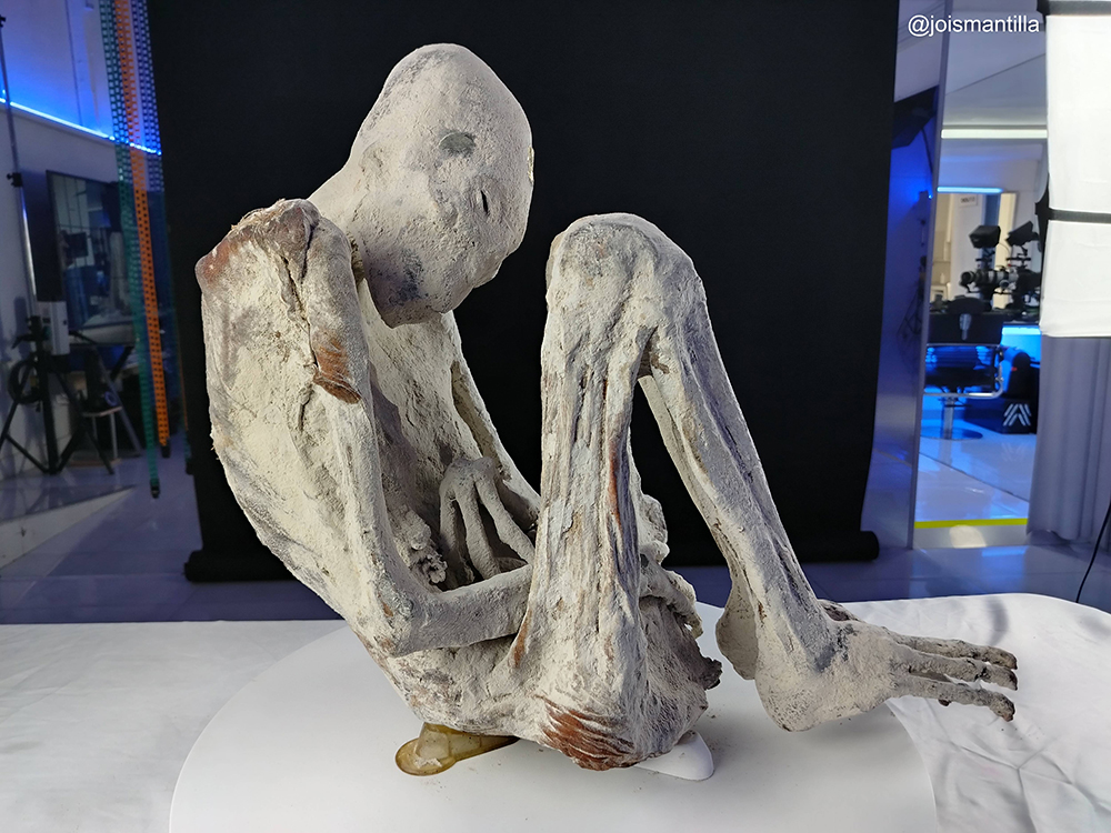
Key Findings
| Finding | Description |
|---|---|
| Complex craniofacial anomalies | Smooth skull enlargement, absence of sagittal suture, enlarged orbits, sloping facial bones, and specific dental configurations |
| Distinct anatomical differences | Thinner cranial bones, smaller cranial base, and significant differences in skull shape compared to modern humans |
| Reduced cranial volume | Approximately 18% smaller cranial capacity than modern humans, with smaller frontal compartment and posterior fossa |
| Advanced imaging techniques | CT reconstruction and digital virtual endocasts provide precise measurements surpassing traditional anthropometric methods |
| Broader contextual anomalies | Elongated skull studied alongside tridactyl features, absence of ear pinnae, slanting eyelids, and metallic implants |
Research Document
Cranial Studies Montserrat
Main Takeaways
- Complex Craniofacial Anomalies: The elongated skull of 'Montserrat' cannot be explained by artificial cranial deformation due to its complex features, such as smooth skull enlargement, absence of sagittal suture, enlarged orbits, sloping facial bones, and specific dental configurations, suggesting a genetic abnormality as a more likely cause.
- Distinct Anatomical Differences: Side-by-side comparisons of 3D CT skull models of 'Montserrat' and a modern human reveal significant differences in skull shape, bone thickness, and facial features, including thinner cranial bones and a smaller cranial base in 'Montserrat'.
- Reduced Cranial Volume: Virtual endocasts show that 'Montserrat's cranial capacity is approximately 18% smaller than that of a modern human, with a smaller frontal compartment and posterior fossa, challenging assumptions about skull size and function.
- Advanced Imaging Techniques: The study utilizes advanced CT reconstruction and digital virtual endocasts to accurately measure cranial volume and depict internal skull structures, surpassing traditional anthropometric methods in precision.
- Broader Contextual Anomalies: The elongated skull must be studied in the context of other anomalies in 'Montserrat', such as tridactyl hands and feet, absence of ear pinnae, slanting eyelids, and metallic implants, indicating a unique biological and possibly cultural significance.