
Switch language
Estudios Craneales
Reconstrucción 3D por TC y análisis de endocastos craneales virtuales digitales
About the Doctor
Dr. K. H. Fung, radiólogo y especialista en visualización médica 3D.
- Radiólogo jubilado con más de 40 años de experiencia en radiología diagnóstica.
- Intereses especiales: visualización médica 3D, radiología intervencionista y neurointervención.
- Artista en la intersección de arte y ciencia, utilizando experiencia en visualización médica 3D.
- Co-ganador del primer lugar en el Desafío Internacional de Visualización Científica e Ingeniería 2007 organizado por la revista Science y la National Science Foundation (EE.UU.).
- Obras de arte exhibidas en museos en EE.UU., Europa, China, Australia y Hong Kong.
- Exposiciones actuales en 2025: Museo de Ciencias Médicas de Hong Kong (imágenes médicas 3D y 4D estereoscópicas y arte) y Nina Park (colección de fósiles de madera Nina) en Hong Kong.
- Colabora con Science Photo Library; obras de arte disponibles en medios digitales internacionales, revistas, libros y journals.
Este estudio arroja luz sobre la anatomía externa e interna de los cráneos alargados utilizando varias técnicas avanzadas de reconstrucción y segmentación por TC, incluyendo endocastos craneales virtuales digitales con comparaciones con cráneos humanos modernos.
Resumen del Estudio
Current Specimen: Montserrat
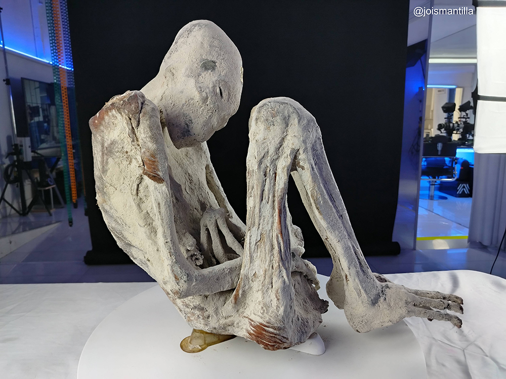
Este estudio se centra en el cráneo alargado de 'Montserrat', examinando tanto la anatomía externa como interna utilizando técnicas avanzadas de reconstrucción por TC y endocastos craneales virtuales digitales.
Hallazgos Clave
| Hallazgo | Descripción |
|---|---|
| Anomalías craneofaciales complejas | Agrandamiento suave del cráneo, ausencia de sutura sagital, órbitas agrandadas, huesos faciales inclinados y configuraciones dentales específicas |
| Diferencias anatómicas distintivas | Huesos craneales más delgados, base craneal más pequeña y diferencias significativas en la forma del cráneo en comparación con humanos modernos |
| Volumen craneal reducido | Capacidad craneal aproximadamente un 18% menor que los humanos modernos, con compartimento frontal y fosa posterior más pequeños |
| Técnicas de imagen avanzadas | Reconstrucción por TC y endocastos virtuales digitales proporcionan mediciones precisas que superan los métodos antropométricos tradicionales |
| Anomalías en contexto más amplio | Cráneo alargado estudiado junto con características tridáctilas, ausencia de pabellones auriculares, párpados inclinados e implantes metálicos |
Documento de Investigación
Estudios Craneales Montserrat
Conclusiones Principales
- Anomalías Craneofaciales Complejas: El cráneo alargado de 'Montserrat' no puede explicarse por deformación craneal artificial debido a sus características complejas, como el agrandamiento suave del cráneo, la ausencia de sutura sagital, órbitas agrandadas, huesos faciales inclinados y configuraciones dentales específicas, sugiriendo una anormalidad genética como causa más probable.
- Diferencias Anatómicas Distintivas: Comparaciones lado a lado de modelos de cráneos en 3D por tomografía computada de 'Montserrat' y un humano moderno revelan diferencias significativas en la forma del cráneo, grosor de los huesos y características faciales, incluyendo huesos craneales más delgados y una base craneal más pequeña en 'Montserrat'.
- Volumen Craneal Reducido: Los endocastos virtuales muestran que la capacidad craneal de 'Montserrat' es aproximadamente un 18% menor que la de un humano moderno, con un compartimento frontal y una fosa posterior más pequeños, desafiando suposiciones sobre el tamaño y la función del cráneo.
- Técnicas de Imagen Avanzadas: El estudio utiliza reconstrucción avanzada por tomografía computada y endocastos virtuales digitales para medir con precisión el volumen craneal y representar estructuras internas del cráneo, superando en precisión a los métodos antropométricos tradicionales.
- Anomalías en un Contexto Más Amplio: El cráneo alargado debe estudiarse en el contexto de otras anomalías en 'Montserrat', como manos y pies tridáctilos, ausencia de pabellones auriculares, párpados inclinados e implantes metálicos, indicando una importancia biológica y posiblemente cultural única.